Submitted by Christina Rozeik on Fri, 17/12/2021 - 21:32
The most comprehensive cell atlas to date of the human uterus has identified two new epithelial cell states that can be used to distinguish two forms of uterine cancer. Researchers from the Wellcome Sanger Institute, the University of Cambridge and their collaborators also identified the genetic pathways that determine two main endometrial cell types.
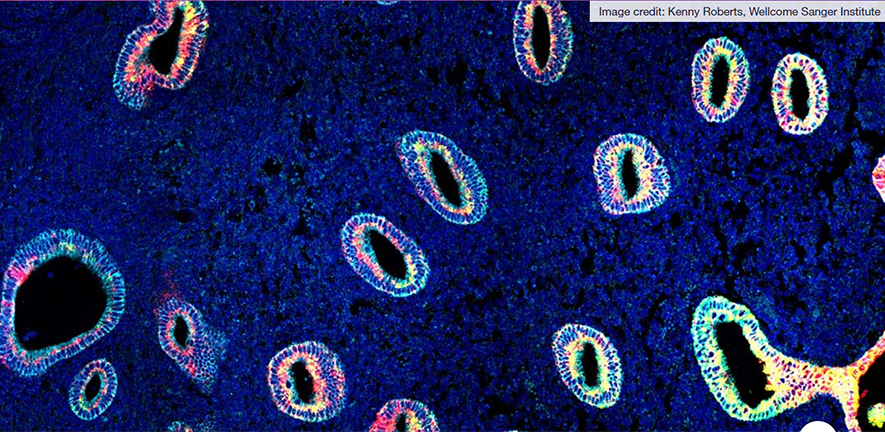
The study, published recently in Nature Genetics, is the first reference map of the uterus to combine single cell and spatial transcriptomics, providing detailed descriptions of cell types as well as where they are situated within the womb lining. Part of the Human Cell Atlas initiative to map every cell type in the human body, the uterine atlas is an important first step towards understanding the endometrium in both health and disease.
The new cell types are found in the endometrium, which is the inner part of the uterus, more commonly known as the womb lining. The top layer of the endometrium sheds and regenerates during a woman’s menstrual cycle, a process regulated by the ovarian hormones oestrogen and progesterone.
In this study, researchers analysed uterine samples from 15 women of reproductive age using single-cell and spatial transcriptomics. This allowed them to generate a cellular map of the human endometrium that accounted for the dynamic changes during the menstrual cycle.
Dr Roser Vento-Tormo, a senior author of the study, said, “We have discovered two new epithelial cell states, and revealed that enrichment of the SOX9+LGR5+ cell population is associated with later stage cancers that pose a greater threat to the patient, as compared to tumours with higher levels of SOX9+LGR5-cells. Understanding the type and severity of a cancer can be crucial in deciding the best course of treatment.”
To further dissect the molecular mechanisms involved in epithelial differentiation, the researchers used endometrial organoids, which were developed by the University of Cambridge. These organoids responded to the ovarian hormones oestrogen and progesterone, mimicking how endometrial tissues function in the body.
The work was carried out as part of the global Human Cell Atlas (HCA) consortium, which aims to create reference maps of all human cell types to understand health and disease.
Reference
Luz Garcia-Alonso, Louis-François Handfield, Kenny Roberts and Konstantina Nikolakopoulou et al., ‘Mapping the temporal and spatial dynamics of the human endometrium in vivo and in vitro’, Nature Genetics 53: 1698–1711. DOI: 10.1038/s41588-021-00972-2.
Adapted from a press release by the Wellcome Sanger Institute. The full version can be found at: https://www.sanger.ac.uk/news_item/uterus-study-is-important-step-towards-understanding-diseases-that-affect-one-third-of-women/.

